Home » Without Label » Diagram Of Shoulder - Anatomy Of The Shoulder Ut Health San Antonio / The anterior deltoid is located on the front of your shoulder.
Diagram Of Shoulder - Anatomy Of The Shoulder Ut Health San Antonio / The anterior deltoid is located on the front of your shoulder.
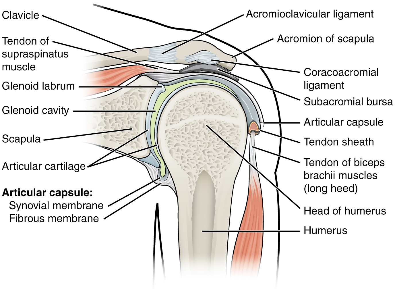
Diagram Of Shoulder - Anatomy Of The Shoulder Ut Health San Antonio / The anterior deltoid is located on the front of your shoulder.. Parts of the right shoulder blade: The shoulder muscles and shoulder tendons involved with shoulder mobility include the four rotator cuff muscle and tendon pairs: The acromioclavicular joint is where the acromion, part of the shoulder blade (scapula) and the collar bone (clavicle) meet. The acromioclavicular joint is formed by an articulation between the lateral end of the clavicle and the acromion process of the scapula. The glenohumeral joint is a joint where the greater tubercle (humeral head at the top of the arm bone) meets the shoulder socket of the scapula, called the glenoid cavity or glenoid fossa.
The shoulder joint (glenohumeral joint) is a ball and socket joint between the scapula and the humerus.it is the major joint connecting the upper limb to the trunk. This is the smallest rotator cuff muscle. The shoulder muscles and shoulder tendons involved with shoulder mobility include the four rotator cuff muscle and tendon pairs: The rotator cuff is a group of 4 principal muscles that stabilize and support the shoulder joint. Its main job is to assist with rotation of the arm away from the body.
Shoulder Muscle Diagram Labeled Dream To Teach from www.dreamtoteach.com The supraspinatus is located on the upper part of the shoulder joint and is involved in abduction (arm raising). The anatomy of the shoulder. The shoulder joint is composed of the glenoid (the shallow shoulder socket) and the head of the upper arm bone known as the humerus (the ball). To further reinforce the shoulder, the four muscles of the rotator cuff extend from the scapula and surround the head of the humerus to both rotate the arm and prevent dislocation. Related posts of diagram of shoulder muscles and tendons muscle anatomy definition. While seated or standing, lift the sore arm forward and to the side about thirty to 45 degrees. The shoulder is made up of two joints, the acromioclavicular joint and the glenohumeral joint. The anterior deltoid is located on the front of your shoulder.
The anterior deltoid is located on the front of your shoulder.
Beyond this, there is also a shoulder joint arrayed in a ball and socket formation, a rotator cuff, and various muscles like the deltoid muscle and the teres major muscle. There are 10 muscles and 11 shoulder tendons related to shoulder mobility. The labrum is a rim of cartilage that surrounds the socket of the shoulder joint. This is the smallest rotator cuff muscle. The glenohumeral joint is a joint where the greater tubercle (humeral head at the top of the arm bone) meets the shoulder socket of the scapula, called the glenoid cavity or glenoid fossa. It is an extremely mobile joint, in which stability has been sacrificed for mobility. The acromioclavicular joint is formed by an articulation between the lateral end of the clavicle and the acromion process of the scapula. The shoulder joint is protected superiorly by an arch, which is formed by the coracoid process of the scapula, the acromion process of the scapula and the clavicle. The labrum also serves as the attachment of a major tendon in the shoulder, the biceps tendon. The bones of the pectoral girdle (clavicle and scapula) provide increased mobility to the. It is the part of the shoulder that borders the chest muscles.its main function is shoulder flexion, which is characterized by raising your upper arms up to the front and overhead.this muscle is targeted during front raises and pressing exercises (e.g. Muscle anatomy definition 12 photos of the muscle anatomy definition muscle belly definition anatomy, muscle fiber anatomy definition, muscle tissue definition anatomy, muscle tissue smooth definition, sternocleidomastoid muscle definition anatomy, human muscles, muscle belly definition anatomy, muscle. The anterior deltoid is located on the front of your shoulder.
These muscles form the outer shape of the shoulder and underarm. This is the smallest rotator cuff muscle. It is the part of the shoulder that borders the chest muscles.its main function is shoulder flexion, which is characterized by raising your upper arms up to the front and overhead.this muscle is targeted during front raises and pressing exercises (e.g. Related posts of diagram of shoulder muscles and tendons muscle anatomy definition. What are common rotator cuff injuries?
The Mechanism Of Shoulder Pain In Aquatics Fina Learning Platform from learning.fina.org The anatomy of the shoulder. The glenohumeral joint, the acromioclavicular joint (a/c joint) and the sternoclavicular joint. Smartdraw includes 1000s of professional healthcare and anatomy chart templates that you can modify and make your own. The glenohumeral joint is a joint where the greater tubercle (humeral head at the top of the arm bone) meets the shoulder socket of the scapula, called the glenoid cavity or glenoid fossa. Muscle anatomy definition 12 photos of the muscle anatomy definition muscle belly definition anatomy, muscle fiber anatomy definition, muscle tissue definition anatomy, muscle tissue smooth definition, sternocleidomastoid muscle definition anatomy, human muscles, muscle belly definition anatomy, muscle. The shoulder joint is composed of the glenoid (the shallow shoulder socket) and the head of the upper arm bone known as the humerus (the ball). It is an extremely mobile joint, in which stability has been sacrificed for mobility. The bones of the pectoral girdle (clavicle and scapula) provide increased mobility to the.
The glenohumeral joint is a joint where the greater tubercle (humeral head at the top of the arm bone) meets the shoulder socket of the scapula, called the glenoid cavity or glenoid fossa.
In this episode of eorthopodtv, orthopaedic surgeon randale c. There are 10 muscles and 11 shoulder tendons related to shoulder mobility. Beyond this, there is also a shoulder joint arrayed in a ball and socket formation, a rotator cuff, and various muscles like the deltoid muscle and the teres major muscle. The shoulder joint is formed where the humerus (upper arm bone) fits into the scapula (shoulder blade), like a ball and socket. It is a flat, gliding joint. It is the part of the shoulder that borders the chest muscles.its main function is shoulder flexion, which is characterized by raising your upper arms up to the front and overhead.this muscle is targeted during front raises and pressing exercises (e.g. What are common rotator cuff injuries? Common rotator cuff injuries include rotator cuff tendonitis and rotator cuff strain, which is a partial or complete tear of the rotator cuff. The four muscles, and their attached tendons that comprise the rotator cuff are the supraspinatus, infraspinatus, subscapularis, and teres minor, and any of these could be where we could find rotator cuff tears. The glenohumeral joint is a joint where the greater tubercle (humeral head at the top of the arm bone) meets the shoulder socket of the scapula, called the glenoid cavity or glenoid fossa. The glenohumeral joint is where the ball (humeral head) and the socket (the glenoid) meet. The muscles of the shoulder support and produce the movements of the shoulder girdle.they attach the appendicular skeleton of the upper limb to the axial skeleton of the trunk. The labrum is a rim of cartilage that surrounds the socket of the shoulder joint.
The supraspinatus, the infraspinatus, the teres minor and the subscapularis. Smartdraw includes 1000s of professional healthcare and anatomy chart templates that you can modify and make your own. In this episode of eorthopodtv, orthopaedic surgeon randale c. These are located in the shoulder blade area, and each related tendon also attaches to the humerus. Image via lh4.googleusercontent.com you can see in the shoulder muscle diagrams that the shoulder is one of the largest and most complex joints in the body.
File 914 Shoulder Joint Jpg Wikimedia Commons from upload.wikimedia.org Numerous muscles help stabilize the three joints of. This is the main muscle that lets you rotate and extend your shoulder. The labrum also serves as the attachment of a major tendon in the shoulder, the biceps tendon. In the case of the these lymph nodes, the nodes serve as a collection point for lymph. The supraspinatus is located on the upper part of the shoulder joint and is involved in abduction (arm raising). The rotator cuff is a group of 4 principal muscles that stabilize and support the shoulder joint. The components of the ball and cup are reversed on the right—a reverse shoulder replacement. The shoulder joint is protected superiorly by an arch, which is formed by the coracoid process of the scapula, the acromion process of the scapula and the clavicle.
The anterior deltoid is located on the front of your shoulder.
The shoulder isn't just one bone, it's actually made up of three different bones and various tendons, ligaments, and muscles.the three bones located in the shoulder are the humerus, the scapula, and the clavicle. The supraspinatus, the infraspinatus, the teres minor and the subscapularis. Muscle anatomy definition 12 photos of the muscle anatomy definition muscle belly definition anatomy, muscle fiber anatomy definition, muscle tissue definition anatomy, muscle tissue smooth definition, sternocleidomastoid muscle definition anatomy, human muscles, muscle belly definition anatomy, muscle. The supraspinatus is located on the upper part of the shoulder joint and is involved in abduction (arm raising). In the case of the these lymph nodes, the nodes serve as a collection point for lymph. Related posts of diagram of shoulder muscles and tendons muscle anatomy definition. The muscles of the shoulder support and produce the movements of the shoulder girdle.they attach the appendicular skeleton of the upper limb to the axial skeleton of the trunk. The shoulder joint (glenohumeral joint) is a ball and socket joint between the scapula and the humerus.it is the major joint connecting the upper limb to the trunk. In this episode of eorthopodtv, orthopaedic surgeon randale c. While seated or standing, lift the sore arm forward and to the side about thirty to 45 degrees. Beyond this, there is also a shoulder joint arrayed in a ball and socket formation, a rotator cuff, and various muscles like the deltoid muscle and the teres major muscle. The shoulder is made up of two joints, the acromioclavicular joint and the glenohumeral joint. All of the nerves that travel down the arm pass through the axilla (the armpit) just under the shoulder joint and are known as the brachial plexus before dividing into the individual nerves.these nerves carry the signals from the brain to the muscles that move the arm.